Uterine anomalies affect the female reproductive system and are usually congenital. Uterine anomalies are differences in the size, division, shape or placement of the uterus.
How is The Uterus Formed?
During the development of the embryo, the zygote, that is, the fertilized egg cell, multiplies by division. As a result of these divisions, the embryo is formed. In the third week of the embryo, three main embryonic layers develop: ectoderm, mesoderm and endoderm. The uterus is derived from part of the mesoderm layer. Mullerian ducts lying on the lateral side of the embryo are formed by cells in the mesoderm layer. Mullerian ducts form the basic building blocks of the embryo's reproductive system. These channels develop and take part in shaping the reproductive organs such as the fallopian tubes, vagina and uterus. Mullerian ducts converge in the middle of the embryo and form an implicit type. This tube is the structure that makes up the uterus. During the developmental stage, other genital structures develop and the female reproductive system is formed. After birth, the uterus continues to grow.


What are Uterine Anomalies?
Uterine anomalies refer to structurally abnormal conditions of the uterus. While the uterus is forming, some people develop uterine anomalies. Uterine anomalies can be listed as follows:
- Shape anomalies; The uterus can develop differently. For example, in a bicornuate uterus, the upper part of the uterus has two separate cavities. In case of double rupture, there are two separate uterine cavities and two cervixes.
- Dimensional anomalies; It is seen as the uterus being smaller or larger than its normal size.
- Septum uterus; It is the presence of one or more sections called septum in the inner part of the uterus. The uterine cavity can be partially or completely divided.
- Unicornuate uterus; The presence of a structure reduced in half on only one side of the uterus. On one side is the normal uterine cavity and on the other is a small cavity.
- Intrauterine adhesions (Asherman syndrome); Adhesions may occur in the inner layer of the uterus after surgical interventions and delivery. This adhesion can cause infertility, menstrual irregularities and pregnancy complications.
Uterine anomalies are usually congenital and are recognized in adolescence. However, in some people, uterine anomaly can be noticed during pregnancy or after birth.
What Causes Uterine Anomaly?
Uterine anomaly can cause many different problems. Some of these problems are as follows:
- Fertility issues; Some anomalies prevent the egg from reaching the uterus and the fertilized egg from implanting in the uterus. Experiencing this situation reduces the chance of pregnancy.
- Recurrent miscarriages; Experiencing uterine anomalies increases the risk of miscarriage. Pregnancy results in miscarriage as a result of the uterus failing to provide adequate support.
- Ectopic pregnancy; It is a condition in which a fertilized egg implants in the uterine cavity outside of the uterus. Urgent medical attention is required.
- Premature birth; Premature birth may occur as a result of the uterus not providing the necessary support in the early stages of pregnancy. This can cause health problems in babies.
- Painful menstrual period; Uterine anomalies, a condition that affects the normal functions of the uterus, can cause severe pain during menstruation.
- Infertility
- Pregnancy complications


What Causes Uterine Anomalies?
The reason for the formation of uterine anomalies is complex. The cause of some conditions may not be fully understood. It usually occurs during the development of the uterus and other reproductive organs during the embryonic period. While the embryo is forming, the uterus and other reproductive organs are formed. The causes of uterine anomalies are as follows:
- Genetic factors
- Environmental effects
- Abnormalities in the development of Muller's duct
- Problems occurring in the vascular process
- Postpartum factors
Detection of Uterine Deformities
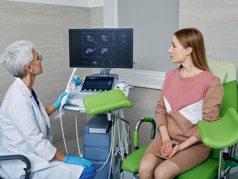
Imaging methods are used to detect deformities of the uterus. Ultrasonography, Magnetic Resonance Imaging (MRI), Hysterosalpingography (HSG) and Hysteroscopy methods can be used. Hysterosalpingography provides X-ray imaging of the uterus and fallopian tubes by injecting contrast material into the uterus. Hysteroscopy is the name given to the endoscopic method used to examine the inside of the uterus. Uterine deformities are usually detected by these methods and physical examination.
Treatment of Uterine Anomalies
Treatment of uterine anomalies is provided according to what the abnormality is and its severity. Some uterine anomalies do not show any symptoms. For this reason, it can be difficult to understand at an early stage. Treatment options for common uterine anomalies are as follows:
- Septate uterus; With hysteroscopic surgery, the septum is removed from the uterus. It is aimed to correct the structure of the uterus and is performed in a minimally invasive way.
- bicornuate uterus; Surgical intervention may be provided to restore the uterus to its normal shape. Hysteroscopy or open surgery can be used.
- unicornuate uterus; Since one side is normal, pregnancy can occur on one side. Therefore, it may not require treatment. However, it should be followed regularly.
- Intrauterine adhesions; Adhesions can be opened with hysteroscopic surgery.
- Uterine size anomalies; This anomaly, which usually does not show symptoms, does not require treatment.
Hysteroscopic surgery is performed from the vaginal area and does not require any incisions.
Hysteroscopy (HSG), magnetic resonance imaging (MRI) or 3D ultrasonography is performed to diagnose uterine malformation. A normal uterus is shaped like an inverted pear. The cervix is cylindrical and has a 2-3 cm long cervix. Uterus that is much less developed than normal means underdeveloped uterus. This can lead to inability to conceive. Having a unilateral uterus means having one fallopian tube instead of two. Half of the uterus is formed. Uterine checking is done with a speculum examination of the vagina and cervix. Manual examination gives an idea about the ovaries and uterus. For a more detailed check, a vaginal ultrasound may be required.How is uterine malformation diagnosed?
What does a healthy uterus look like?
What does underdeveloped uterus mean?
What does a unilateral uterus mean?
How is the uterus checked?


